|
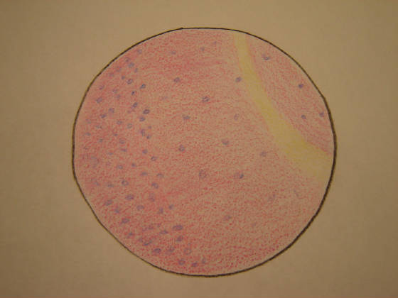
The cerebellum is a part of the brain located just above the brainstem towards the back of the
head. It is divided into two parts: the cerebellar cortex and the deep cerebellar nuclei. The cerebellar cortex is comprised
of three layers. The outermost layer, called the molecular layer, is composed of the stellate and basket cells which are two
types of inhibitory interneurons. The middle layer is called the Purkinje Cell Layer, which gets its name from the Purkinje
cells which are found there. The Purkinje cells send inhibitory messages to the deep cerebellar nuclei. Each of these cells
receives synapses (impulses) from roughly 100 000 axons of the granule cells from the cerebellar cortex. The innermost layer
is called a granular layer, which is made up of granular cells that sense impulses from outside the cerebellum and sent outputs
to other cells inside the cerebellum. The directions for the cerebellar functions are sent from the primary motor cortex to
the spinal cord, which are then received by the cerebellum from the cerebral cortex, visual cortex and somatosensory cortex.
The cerebellum is partitioned into the anterior lobe, the posterior lobe and the flocculonodular lobe, between which the primary
and posterior lateral fissures run. Although the cerebellum appears small, it contains half of the neurons in the brain and
is responsible for guiding movement based on sensory feedback (mostly visual), directing attention, measuring time and other
cognitive operations
|