|
Male mammals, including humans, have two testes (or testicles) which are components of the reproductive
and endocrine systems. The testes are responsible for producing sperm and male sex hormones. Both roles are controlled by
Luteinizing Hormone (LH) and Follicle-Stimulating Hormone (FSH). Each testis is ovular and measures roughly 5cm long and 3cm
in diameter. The testes are located in an external pouch called a scrotum, which hangs just behind the penis. Despite their
external location, the testes are developed internally, and descend through the inguinal canal shortly before or after birth.
This external location provides the testes with a temperature about 3° C lower than normal body temperature,
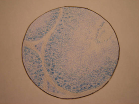
which is ideal for the production of healthy sperm. Surrounding each testis is a connective tissue which extends upwards and
divides the testis onto approximately 250 lobules. Inside each lobule are 1 to 4 semniferous tubules, tightly coiled and lined
with cells which produce sperm cells. The semniferous tubules lead to the epididymis where the sperm cells mature and move
to the vas deferens. Upon sexual arousal, the sperm move through an ejaculatory duct and the prostate ejaculates the sperm
through muscular contractions.
|