|
The trachea, also known as a windpipe in, is a strong and flexible tube comprised of a layer of
connective and muscular tissue which forms the airway in mammals. It has a diameter of about 1.5 cm and length of 10 cm. The
larynx is located at the upper end of the trachea, which runs to the upper part of the chest and branches into tracheoles.
The tracheoles extend into the left and right bronchi, which enter the lungs via bronchial tubes. The trachea is lined with
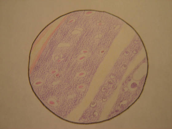
ciliated cells and supported by cartilage rings.These rings of cartilage are u-shaped and roughly 16 to 20 in number, depending
on age and sex. Goblet cells in the trachea secrete a mucous which acts to trap dust and other foreign particles. The beating
of the cilia on the ciliated cells moves the trapped particles away from the lungs and towards the nose or mouth where they
are expelled.
|