|
The stomach is located on the left side of the abdominal cavity, between the esophagus and the duodenum
in humans. It is an organ used primarily for the digestion of food, although absorption of nutrients does not normally occur
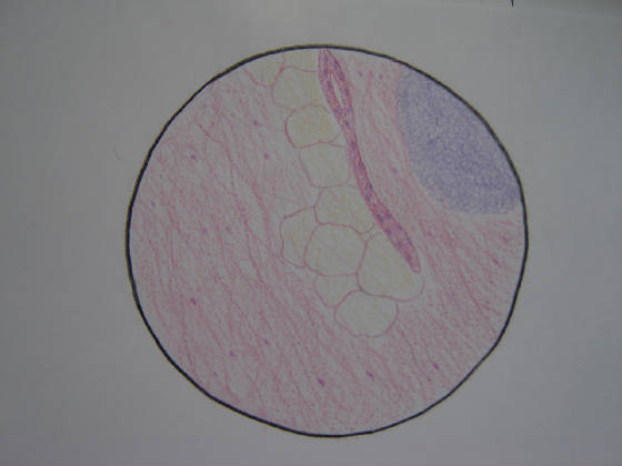
there. The secretion of hydrochloric acid maintains an acidic environment within the stomach, which is divided into five sections,
each with different cells of different functions. The stomach walls consist of a number of layers, and different cells within
these layers are responsible for the production of pepsinogen, hydrochloric acid, and mucous. Pepsinogen is the inactive form
of pepsin, which breaks down proteins; hydrochloric acid kills most bacteria in food, promotes appetite and activates pepsin
from pepsinogen; and mucous helps protect the stomach from self-digestion. The innermost layer controls the motion of the
stomach that physically breaks down the food (called chyme during digestion). This chyme moves from the stomach to the first
part of the small intestine, known as the duodenum, through the pyloric sphincter. The duodenum is a slightly acidic, hollow
junction between the stomach and the jejunum (the second part of the small intestine). It measures roughly 18cm in length
and contains two important ducts, the bile and pancreatic ducts.
|